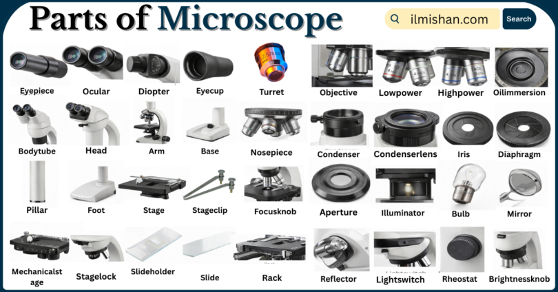
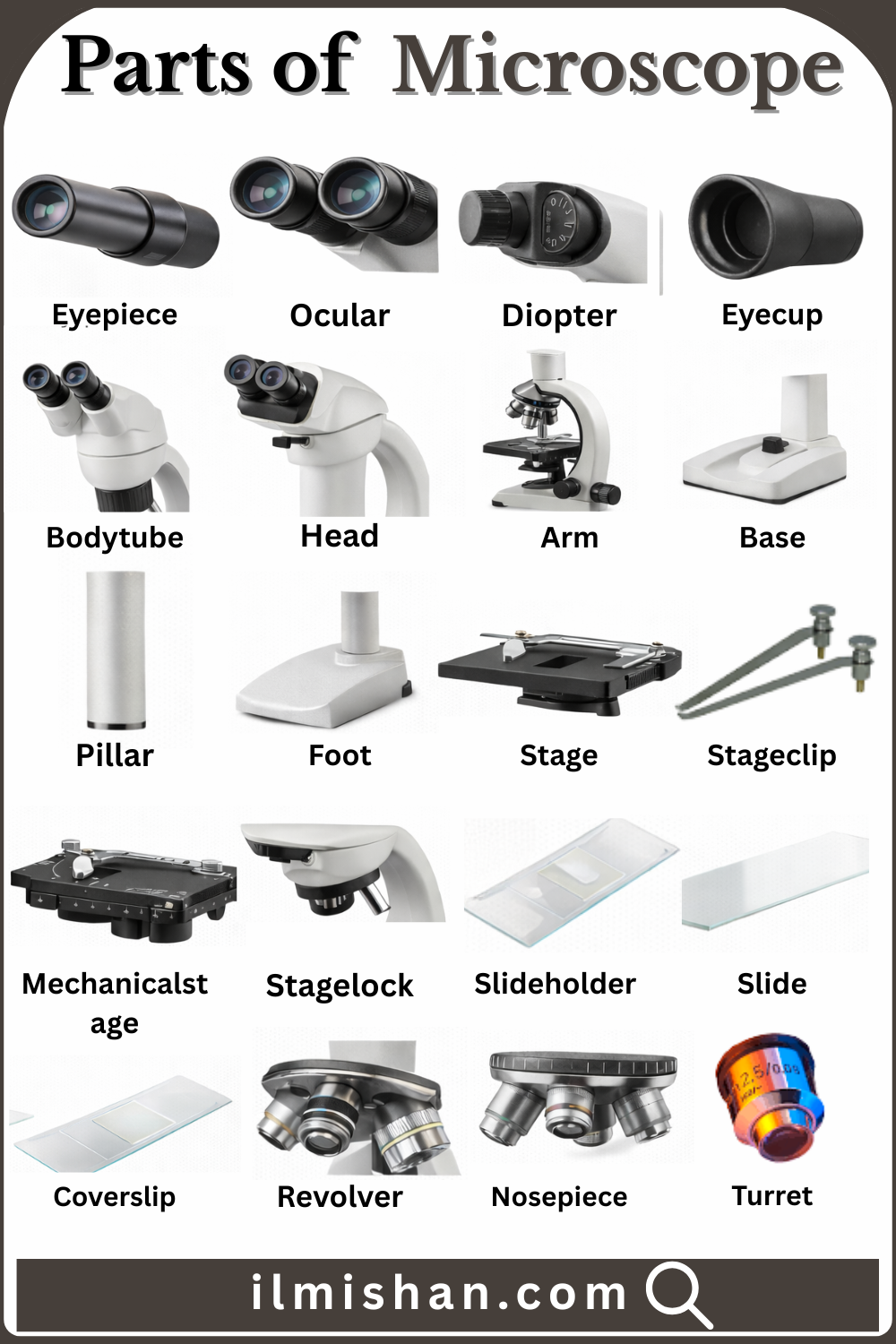
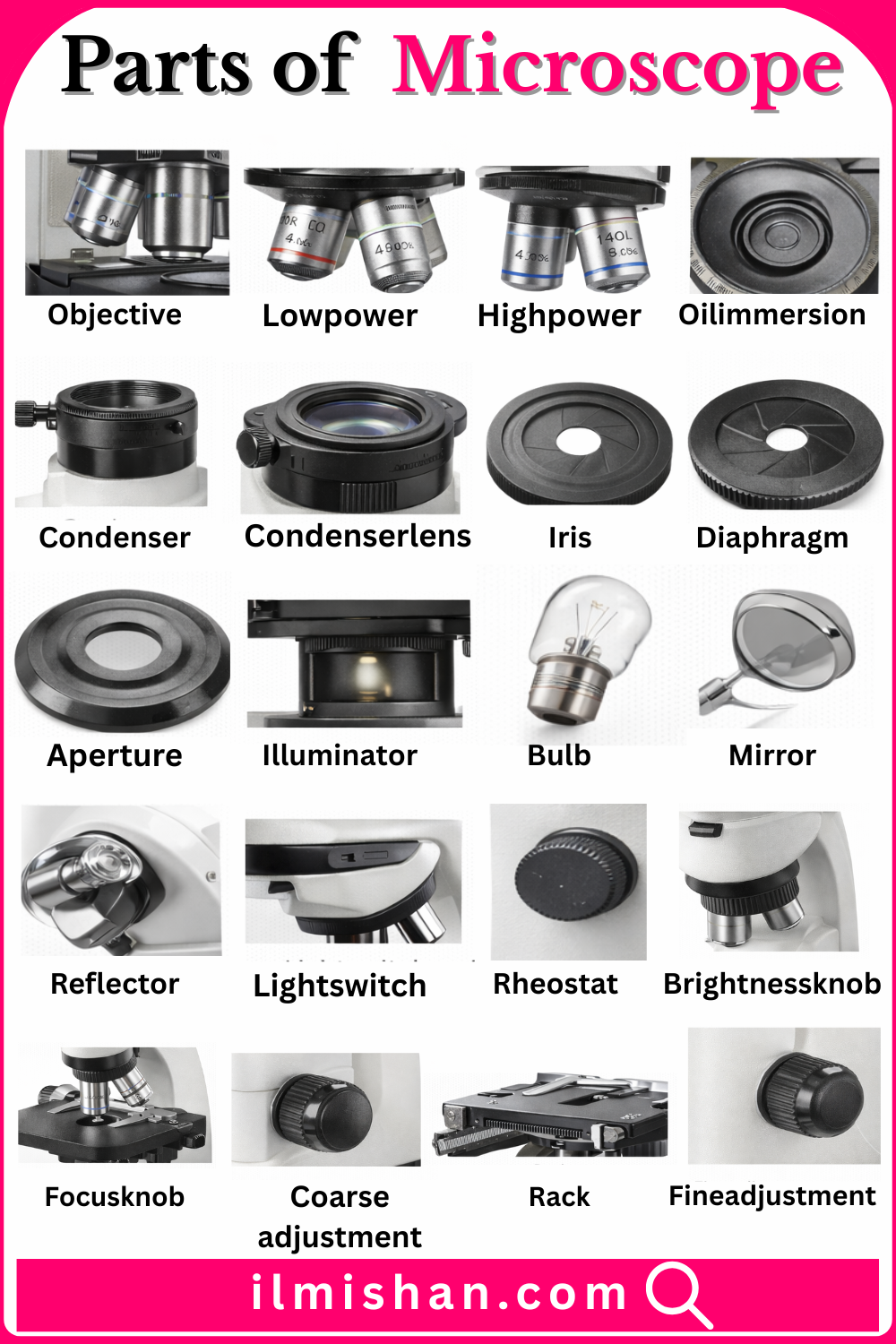
Parts of Microscope with Names list
- Eyepiece
- Ocular
- Diopter
- Eyecup
- Bodytube
- Head
- Arm
- Base
- Pillar
- Foot
- Stage
- Stageclip
- Mechanicalstage
- Stagelock
- Slideholder
- Slide
- Coverslip
- Revolver
- Nosepiece
- Turret
Microscope Parts Names in English with there Pictures
- Eyepiece
Lens located at the top of the microscope through which you look to observe the specimen. It magnifies the image formed by the objective lens, making small details appear larger and clearer to the viewer.
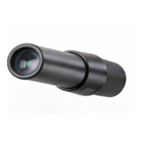
- Ocular
Another term for the eyepiece lens that provides the final level of magnification. It works together with the objective lens to enlarge the specimen’s image for comfortable and detailed viewing.
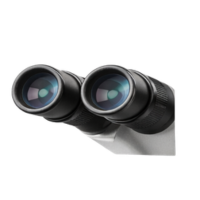
- Diopter
Adjustable ring usually found on one eyepiece that allows users to fine-tune focus according to differences between their eyes, ensuring a sharp and balanced image without eye strain.
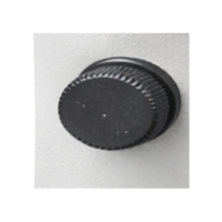
- Eyecup
Soft rubber or plastic covering around the eyepiece that blocks unwanted light from entering the eye and provides comfort during long observation sessions under the microscope.
- Bodytube
Hollow tube connecting the eyepiece to the objective lenses. It keeps optical components properly aligned so light travels correctly, ensuring clear magnification and accurate image formation.
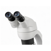
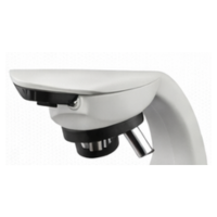
- Head
Upper section of the microscope that holds the eyepiece and connects to the nosepiece. It supports optical parts and directs the image from objectives to the viewer’s eye.
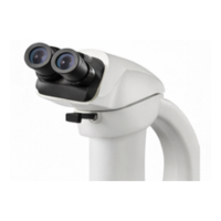
- Arm
Strong curved support connecting the head to the base. It holds major components together and is the correct part to grip when carrying the microscope safely.
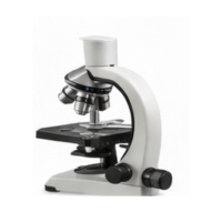
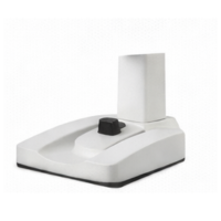
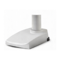
- Base
Heavy bottom platform of the microscope that provides stability and balance. It supports the entire structure and often houses the built-in light source or electrical components.
- Pillar
Vertical supporting structure rising from the base that strengthens the frame and helps hold the arm and stage assembly firmly in position during use.
- Foot
Lower supporting portion of the base that rests directly on the working surface, preventing slipping and maintaining steady positioning while observing specimens.
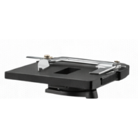
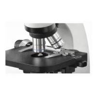
- Stage
Flat platform where the microscope slide is placed. It contains a central opening to allow light to pass through the specimen for clear examination.
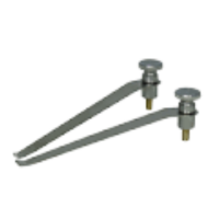
- Stageclip
Metal clip attached to the stage that holds the slide firmly in place, preventing movement while focusing or adjusting magnification levels.
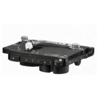
- Mechanicalstage
Advanced stage mechanism equipped with control knobs that move the slide precisely left, right, forward, and backward for detailed scanning of the specimen.
- Stagelock
Locking device that secures the stage at a chosen height or position, preventing accidental movement during focusing or transportation.
- Slideholder
Component on the stage designed to grip and stabilize the slide securely while allowing smooth, controlled adjustments during observation.
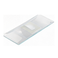
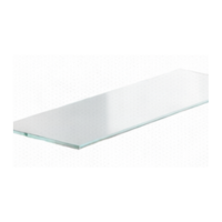
- Slide
Thin rectangular glass plate used to mount and support a specimen sample so it can be examined under the microscope.
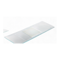
- Coverslip
Small, thin square of glass placed over the specimen on the slide to protect it, flatten it evenly, and improve image clarity.
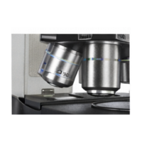
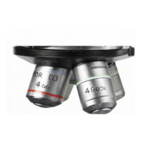
- Revolver
Rotating disc that holds multiple objective lenses. It allows quick switching between different magnification powers by turning it gently.
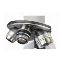
- Nosepiece
Circular rotating part attached below the head that carries objective lenses and positions them accurately over the specimen.
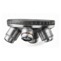
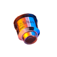
- Turret
Another name for the revolving nosepiece that supports several objective lenses and rotates smoothly to change magnification during observation.
Microscope Parts Names in English with there Pictures
Explore Microscope parts and Name
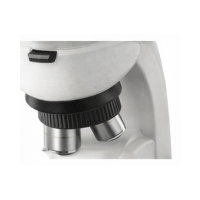
- Objective
- Lowpower
- Highpower
- Oilimmersion
- Condenser
- Condenserlens
- Iris
- Diaphragm
- Aperture
- Illuminator
- Bulb
- Mirror
- Reflector
- Lightswitch
- Rheostat
- Brightnessknob
- Focusknob
- Coarseadjustment
- Fineadjustment
- Rack
Common Parts of Microscope with their Names and Pictures
- Objective
Lens located near the specimen that gathers light and magnifies the image, working with the eyepiece to provide overall magnification.
- Lowpower
Objective lens with low magnification, typically 4x or 10x, used for scanning and locating the specimen.
- Highpower
Objective lens with higher magnification, typically 40x or 50x, used for detailed observation of fine structures.
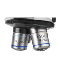
- Oilimmersion
High-magnification lens, usually 100x, used with immersion oil to increase resolution and clarity of tiny specimens.
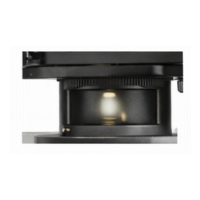
- Condenser
Lens system beneath the stage that focuses light onto the specimen for clear and bright illumination.
- Condenserlens
Individual lens within the condenser that directs and concentrates light onto the specimen to enhance contrast and detail.
- Iris
Adjustable component of the condenser controlling the amount of light passing through the specimen.
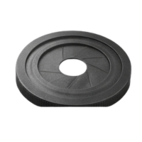
- Diaphragm
Part of the condenser that adjusts light intensity and contrast by opening or closing its aperture.
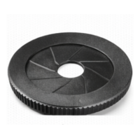
- Aperture
Opening in the diaphragm or condenser allowing light to pass through and illuminate the specimen.
- Illuminator
Light source of the microscope, either built-in or external, that provides illumination for viewing the specimen.
- Bulb
Electric light used in the illuminator to produce a steady and consistent light for observation.
- Mirror
Reflective surface used in older microscopes to direct external light through the condenser and onto the specimen.
- Reflector
Part that redirects light from an external source through the microscope to illuminate the specimen.
- Lightswitch
Switch controlling the microscope’s built-in light source, turning it on or off.
- Rheostat
Control dial that adjusts the intensity of the illuminator light for proper brightness.
- Brightnessknob
Knob used to increase or decrease light intensity for optimal viewing and contrast.
- Focusknob
Knob used to adjust the focus of the specimen, moving the stage or bodytube for clarity.
- Coarseadjustment
Large knob for rapid movement of the stage or bodytube to bring the specimen into rough focus.
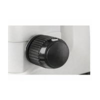
- Fineadjustment
Small knob for precise focusing, allowing detailed and sharp visualization of the specimen.
- Rack
Part of the focusing mechanism or stage assembly that moves the stage vertically to focus the specimen.
Common Parts of Microscope with their Names and Pictures